Skull Anatomy⁚ A Comprehensive Overview
This overview explores the intricate structure of the human skull‚ encompassing cranial and facial bones‚ their sutures and joints‚ developmental processes‚ and clinical significance. Detailed anatomical descriptions and evolutionary perspectives are provided‚ alongside resources for further learning. Understanding skull anatomy is crucial for various medical fields.
Introduction to Skull Anatomy
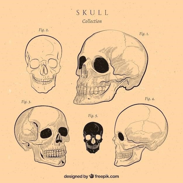
The human skull‚ a complex bony structure‚ serves as the protective casing for the brain and houses the sensory organs of sight‚ hearing‚ smell‚ and taste. Composed of numerous individual bones‚ it’s divided into two main parts⁚ the neurocranium (cranium) protecting the brain‚ and the viscerocranium (facial skeleton) forming the framework of the face. Understanding the skull’s anatomy is crucial in various fields‚ including medicine‚ anthropology‚ and forensic science. This intricate structure‚ formed by intramembranous ossification‚ features numerous sutures (fibrous joints) that fuse in adulthood. The skull’s internal structure includes the cranial fossae‚ which house specific brain regions. Detailed knowledge of skull anatomy requires a thorough understanding of individual bone structures‚ their articulations‚ and developmental processes. The study of the skull also incorporates an evolutionary perspective‚ revealing insights into species adaptations and phylogenetic relationships.
Cranial Bones⁚ Structure and Function
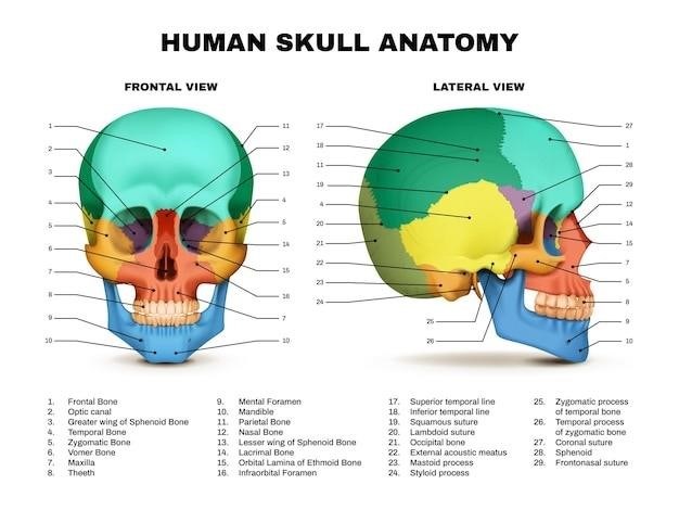
The neurocranium‚ or braincase‚ comprises eight major flat bones⁚ the frontal‚ occipital‚ sphenoid‚ ethmoid‚ and paired parietal and temporal bones. The frontal bone forms the forehead and contributes to the anterior cranial fossa. The occipital bone‚ situated posteriorly‚ forms the foramen magnum‚ through which the spinal cord passes. The sphenoid bone‚ a complex‚ keystone-shaped bone‚ sits at the base of the skull‚ contributing to both the anterior and middle cranial fossae. The ethmoid bone‚ located anterior to the sphenoid‚ forms part of the nasal cavity and orbits. The paired parietal bones form the majority of the skull’s superior and lateral aspects‚ while the paired temporal bones‚ located inferiorly‚ house the inner ear structures and articulate with the mandible. These bones‚ joined by sutures‚ protect the brain and provide attachment points for muscles involved in head movement and facial expression. Their precise structure and arrangement are crucial for brain protection and overall cranial function.
Facial Bones⁚ Structure and Function
The facial skeleton‚ or viscerocranium‚ consists of 14 bones that contribute to the structure of the face‚ nasal cavity‚ and orbits. Paired bones include the maxillae (upper jaw)‚ zygomatic bones (cheekbones)‚ nasal bones (bridge of the nose)‚ lacrimal bones (medial walls of the orbits)‚ palatine bones (posterior hard palate)‚ and inferior nasal conchae (turbinates). Unpaired bones are the mandible (lower jaw)‚ vomer (nasal septum)‚ and hyoid bone (unique in that it does not directly articulate with other bones). The maxillae are central to the facial skeleton‚ forming the upper jaw‚ supporting the teeth‚ and contributing to the hard palate and orbits. Zygomatic bones form the prominent cheekbones and contribute to the orbits. Nasal bones form the bridge of the nose‚ while lacrimal bones contribute to the tear drainage system; The mandible‚ the only movable bone of the skull‚ allows for chewing and speaking. Together‚ these bones provide structural support for the face‚ protect sensory organs‚ and facilitate essential functions such as mastication and respiration.
Sutures and Joints of the Skull
The skull’s unique articulation system‚ comprised primarily of fibrous joints called sutures‚ allows for growth and development during childhood while providing significant structural integrity. These immobile joints are formed by dense connective tissue that interdigitates between adjacent bones. Key sutures include the coronal suture (between frontal and parietal bones)‚ sagittal suture (between parietal bones)‚ lambdoid suture (between parietal and occipital bones)‚ and squamous sutures (between parietal and temporal bones). The sphenoid and ethmoid bones contribute to numerous complex articulations within the skull base. While most sutures fuse completely during adulthood‚ some retain mobility‚ allowing for slight skull movement. The temporomandibular joint (TMJ)‚ a synovial joint connecting the mandible to the temporal bone‚ is a significant exception‚ permitting essential jaw movements for speech and mastication. Understanding the structure and function of these sutures and joints is crucial in diagnosing and treating skull fractures and other craniofacial conditions.
Development of the Skull⁚ Ossification and Growth
Intramembranous ossification‚ a process where bone forms directly from mesenchymal tissue‚ primarily shapes the flat bones of the skull vault (calvaria). This begins with the formation of ossification centers‚ which gradually expand and fuse‚ creating the individual cranial bones. The skull base develops through endochondral ossification‚ a process involving cartilage as an intermediary. Cartilage models are formed first‚ which then undergo ossification to become bone. This dual process results in the complex structure of the adult skull. Growth occurs at the sutures‚ where bone apposition and remodeling take place. The timing and extent of this growth are influenced by genetic factors‚ hormonal regulation‚ and environmental factors. Fontanelles‚ membranous areas between the bones of the fetal skull‚ allow for brain growth and deformation during childbirth. These fontanelles gradually close during infancy‚ completing the skull’s ossification. Understanding this developmental process is key in assessing normal skull growth and identifying potential developmental abnormalities.
Evolutionary Perspective on Skull Anatomy
This section explores the fascinating evolution of the skull across diverse vertebrate lineages‚ highlighting adaptations related to feeding‚ sensory perception‚ and brain development. Comparative analysis reveals key evolutionary transitions and functional significance.
Skull Evolution in Vertebrates
Vertebrate skull evolution showcases a remarkable diversity of forms reflecting adaptation to varied ecological niches and lifestyles. Early vertebrates possessed simple cartilaginous skulls‚ gradually evolving into ossified structures with increasing complexity. The evolution of the jaw‚ a pivotal innovation‚ dramatically altered feeding strategies and impacted skull morphology. Fish skulls‚ particularly in crossopterygians‚ exhibit a movable hinge‚ a feature significant for understanding the transition to terrestrial life. Amphibians show modifications in skull structure associated with their amphibious lifestyle‚ while reptiles exhibit further diversification. Mammalian skulls are characterized by a unique arrangement of bones‚ reflecting their specialized feeding mechanisms and enhanced sensory capabilities. The evolution of the human skull is marked by changes in brain size‚ facial features‚ and dentition‚ influenced by environmental pressures and dietary shifts. Avian skulls display remarkable adaptations for flight‚ including lightweight construction and specialized jaw structures. Understanding vertebrate skull evolution requires a comparative approach‚ integrating anatomical‚ paleontological‚ and developmental data.
Comparative Skull Anatomy
Comparative skull anatomy reveals fascinating insights into evolutionary relationships and adaptations across diverse vertebrate lineages. Analyzing skull structures across species‚ including bone morphology‚ joint types‚ and relative sizes of cranial and facial regions‚ reveals patterns of evolutionary divergence and convergence. For instance‚ comparing the skulls of different mammals highlights variations in dentition reflecting dietary specializations—carnivores‚ herbivores‚ and omnivores exhibit distinct dental adaptations. Similarly‚ comparing bird skulls with those of reptiles reveals modifications associated with flight‚ including reduced weight and specialized jaw structures for efficient feeding. Studying the skulls of extinct species provides crucial evidence for understanding evolutionary transitions and reconstructing phylogenetic relationships. Comparative approaches‚ involving detailed anatomical descriptions and quantitative analyses‚ are essential for interpreting evolutionary pathways and understanding the functional significance of skull morphology. This comparative perspective enhances our understanding of the complex interplay between genotype and phenotype in shaping vertebrate skull diversity.

Clinical Significance of Skull Anatomy
Understanding skull anatomy is paramount in diagnosing and treating head injuries‚ including fractures and associated neurological complications. Surgical procedures involving the skull necessitate precise anatomical knowledge for successful interventions.
Skull Fractures and Injuries
Skull fractures‚ ranging from linear to comminuted‚ are significant clinical concerns. Their diagnosis often involves imaging techniques like X-rays and CT scans to assess the extent of the damage. The location of the fracture influences the potential neurological complications. Basilar skull fractures‚ affecting the base of the cranium‚ can lead to cerebrospinal fluid leakage from the ears or nose‚ a life-threatening condition requiring urgent medical attention. Depressed fractures‚ where bone fragments are pushed inward‚ pose a risk of brain injury. Management strategies vary depending on the fracture type and severity‚ ranging from conservative approaches (observation and pain management) to surgical intervention for displaced or severely compromised fragments. The potential for intracranial hemorrhage‚ brain swelling‚ and infection necessitates careful monitoring and appropriate treatment. Prompt and accurate assessment is crucial to minimize morbidity and mortality.
Surgical Approaches to the Skull
Surgical access to the skull necessitates a thorough understanding of its complex anatomy. Various approaches exist‚ each chosen based on the location and nature of the surgical target. Craniotomies‚ involving the removal of a portion of the skull‚ provide access to the brain for procedures such as tumor resection‚ aneurysm clipping‚ or trauma repair. The choice of craniotomy approach depends on factors including the location of the pathology‚ the extent of surgical exposure required‚ and the surgeon’s preference. Minimally invasive techniques are increasingly employed to reduce surgical trauma‚ including endoscopic-assisted procedures. Preoperative planning often involves 3D imaging for precise surgical navigation and to minimize risk to critical structures like blood vessels and nerves. Postoperative care is essential to manage pain‚ swelling‚ and potential complications‚ ensuring optimal patient recovery.

Resources for Further Learning
Explore comprehensive online resources‚ interactive 3D models‚ and detailed anatomical PDFs for in-depth study of skull anatomy. These resources offer valuable supplementary materials for enhanced learning.
Recommended Skull Anatomy PDFs
Several valuable PDFs offer detailed insights into skull anatomy. These resources often include high-resolution images‚ diagrams‚ and tables summarizing key features of the cranial and facial bones. Look for PDFs that provide clear descriptions of bone structures‚ including foramina‚ processes‚ and sutures. Some PDFs may focus on specific aspects‚ such as the development of the skull or the clinical relevance of skull fractures. Others may offer a more comprehensive overview‚ covering both the macroscopic and microscopic anatomy. Ensure the PDF’s information aligns with current anatomical terminology and understanding. Cross-referencing information across multiple sources is always a good practice to ensure accuracy and a thorough understanding of the subject matter. When selecting a PDF‚ consider the author’s credentials and the publication date to ensure the information is reliable and up-to-date. Many academic institutions and medical organizations provide free or low-cost access to high-quality anatomical resources.
Online Resources and Interactive Models
Beyond PDFs‚ numerous online resources offer interactive learning experiences for studying skull anatomy. Websites and educational platforms provide 3D models allowing for rotation and zooming‚ enabling detailed examination of bone structures from various angles. These interactive models often include labeling functions‚ quizzes‚ and detailed descriptions of each bone and its features. Some websites offer virtual dissection tools‚ simulating the experience of examining a real skull. These resources are particularly helpful for visualizing complex spatial relationships between bones and understanding the three-dimensional structure of the skull. Furthermore‚ many online resources offer videos and animations explaining the development‚ function‚ and clinical significance of different skull components. These dynamic learning tools can significantly enhance comprehension and retention of information compared to static images. Always verify the credibility of online resources by checking the source and ensuring the information aligns with established anatomical knowledge.


